Cerebrovascular Disease
Cerebrovascular disease is treated at UC Davis Health by a multidisciplinary team, consisting of staff from Neurological Surgery, Neuroradiology, Interventional Neuroradiology, Neurology, Pulmonary/Critical Care, Anesthesiology, Nursing, Physical Medicine and Rehabilitation, Physical Therapy, Occupational Therapy, and Speech Therapy. We provide comprehensive treatment to patients with a variety of neurovascular problems.
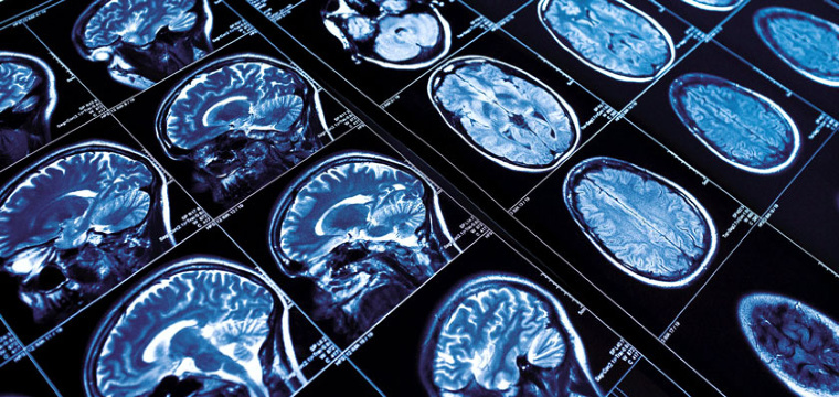
A stroke is an injury to the brain caused by lack of blood flow. It is a common condition affecting more than 750,000 Americans each year. It is the third leading cause of death and the leading cause of adult disability. A stroke can be caused by blockages in the blood vessels that feed the brain (ischemic) or a break in the blood vessels causing bleeding in the brain (hemorrhagic). Ischemic stroke, if recognized early can be treated with medicines given intravenously by special clot busters given directly into the vessels in the brain. This specialized care of ischemic stroke is directed by a stroke neurologist. Hemorrhagic stroke, caused by rupture of a blood vessel feeding the brain often involves the care of a neurosurgeon.
- Weakness or numbness especially on one side of the body
- Difficulty talking or understanding
- Sudden change in vision
- Sudden trouble walking or loss of balance or coordination
- Sudden severe headache with no known cause
Carotid endarterectomy (CEA) is a surgical procedure used to treat narrowing (stenosis) of the carotid artery. The carotid arteries play a vital role in providing oxygen and nutrients to the brain. Narrowing of the carotid arteries can lead to impaired blood flow and stroke. It is more common in people that smoke, have diabetes or hypertension. A landmark research study called the NASCET study examined the question of whether or not patients benefited from surgery to correct a narrowed carotid or if taking medicine would be better. This study suggests that people who have a carotid stenosis greater than 70% and suffer from stroke like symptoms [transient ischemic attacks (TIA)] can reduce the risk of suffering a stroke by 50% if CEA is performed. In contrast, those who have no clinical symptoms and narrowing of less than 70% may not benefit and should consider medical therapy. Carotid endarterectomy is of no proven benefit to occluded vessels.
Your doctor may recommend tests to determine if you have carotid stenosis. One of the first tests is often listening to the neck with a stethoscope. Sometimes a noise is heard called a bruit, which may indicate abnormal blood flow. A second non-invasive test is called carotid ultrasound. This test reliably detects carotid stenosis by measuring blood flow velocities within the carotid artery. A third more definitive test is called an angiogram, used to directly visualize the vessels and measure narrowing. Carotid endarterectomy surgery, to open the blocked vessel, is usually an elective procedure recommended after identifying carotid stenosis in a symptomatic patient. The surgery is performed under anesthesia and requires making an incision on the neck to access the carotid artery. The vessel is then opened to remove the obstruction and restore normal blood flow. The vessel wall is then closed and sutures are placed in the neck. Most patients spend one to two days in the hospital and return home.
Cerebral aneurysms are fairly uncommon, found in only 2% of the population. Aneurysms are abnormal weak spots in the blood vessel wall causing a pouch to form. Because the cerebral aneurysm pouch is weaker than the normal vessel wall, breaks can occur causing a serious condition called subarachnoid hemorrhage. Bleeding in the brain, caused by an aneurysm rupture, is very irritating to the nerves surrounding the brain. The most common symptom is a severe headache often felt to be “the worst headache of my life.” In most cases, this is the first symptom of any problem.
Unruptured aneurysms rarely have any symptoms. In fact, the first symptom of any problem may be an aneurysm rupture, producing some or all of the symptoms listed below:
- Sudden severe headache with no known cause, “worst headache of your life”
- Sensitivity to noise, light
- Neck stiffness
- Weakness, numbness on one side of the body
- Difficulty talking or understanding
- Decreased consciousness
Bleeding in the brain caused by a ruptured aneurysm is usually detectable on standard CT imaging of the brain. For a minority of patients with symptoms of a ruptured aneurysm with normal CT imaging of the brain, a lumbar puncture is helpful in making the diagnosis. Unruptured aneurysms often have no symptoms and are usually found by chance.
At UC Davis, technologically advanced pictures are taken of the aneurysm using a CT scan and contrast to create a three dimensional picture of the blood vessels (CT angiogram). A CT angiogram is a fast, non-invasive alternative to traditional angiography that requires several hours to complete. Sometimes, traditional angiography may be required to learn more about the aneurysm before treating it. These diagnostic studies are used by our doctors to determine where the aneurysm is and how it should be treated.
Not all aneurysms need treatment. Brain aneurysms can be treated by either closing the aneurysm off from the inside (endovascular coiling) or by use of a special clip during brain surgery. Consult with a specialist to find out more about your treatment options.
Aneurysm clipping is a surgical procedure used to correct the abnormal aneurysm pouch and eliminate the risk of rupture. Surgical clipping of cerebral aneurysms has been performed since 1937. This form of aneurysm treatment carries no risk for re-opening of the aneurysm and is accomplished by performing brain surgery to apply a clip directly to the aneurysm to seal it off. A surgical procedure called a craniotomy is performed to visualize the aneurysm. The surgeon will make an incision on the scalp and temporarily remove the bone creating a window into the area of the brain where the abnormal blood vessel is located. The abnormal blood vessel will be repaired using a clip to seal off the abnormal pouch and the bone will be replaced. The scalp incision is then closed with sutures or staples.
Every aneurysm is different. Blood vessel variations make selecting the right treatment a highly individual choice. At UC Davis, a team of expert neurosurgeons and neuroradiologists will review your case to determine which treatment is right for you. In general, aneurysms that have ruptured and are not amenable to coil embolization should be considered for surgical clipping. This reduces the risk of dangerous re-bleeding of the aneurysm which can be fatal.
Patients are typically monitored closely in our specialized neurosurgical intensive care unit (NSICU). Common problems that may arise after aneurysmal SAH include vasospasm, rebleeding, and hydrocephalus.
Standard care includes therapies for the treatment of spasm, once it occurs. There may be other experimental methods of treating spasm, such as new medication to decrease the degree of spasm. As far as we know, there is not any other treatment that attempts to prevent vasospasm from occurring.
We currently are evaluating a promising treatment for patients who have ruptured cerebral aneurysms. A common, harmful complication that patients with blood in the brain develop is a spasm or cramping of the blood vessels in the brain. Although the exact cause of this spasm is unknown, it is believed to be due to muscle constriction and changes of the vessel wall. Both of these cause a narrowing or constriction of the blood vessels in your brain. This constriction of the vessel often does not allow adequate blood flow and oxygen to get to the brain.
Transluminal ballooning is a procedure that opens blood vessels, allowing blood flow and oxygen to get to the brain more easily. We believe that this dilation of the blood vessels lasts for at least 7 days. This procedure is used to treat spasm although it is not commonly used to prevent spasm. We are trying to find out if this procedure, performed immediately after surgery, prevents spasm in the brain and improves patient outcome. Seven other hospitals are participating in this study. The study is funded by the National Institutes of Health (NIH). All patients have been enrolled. We are now evaluating the data we have compiled and hope to have our results published in the near future.
Aneurysms have been found to occur more commonly within familial lines, although they are not inherited. The advent of non-invasive methods of imaging intracranial blood vessels (particularly CT angiogram) has facilitated screening for cerebral aneurysms in people who are at risk. A strong risk factor for cerebral aneurysm, which causes subarachnoid hemorrhage, is a positive family history, defined as two or more first-degree relatives with subarachnoid hemorrhages. The greatest familial risk is associated with an affected sibling. Another strong risk factor is polycystic kidney disease. People with these risk factors are potential candidates for screening.
Please refer to the resources noted on our web page. Resources and information are available online from the Brain Aneurysm Foundation There is a local support group coordinated by our department. Currently the group meets on the forth Saturday of the month from 3-5 pm at the UC Davis Ambulatory Care Center, 4860 Y Street RM 3015 Third Floor.
An arteriovenous malformation is a tangle of vessels located in the brain with an abnormal direct connection between the arteries and the veins. In normal circulation, the “high flow” arteries are separated from “low flow” veins by a capillary bed. Without this important structure, the high flow arteries cause severe dilation in the veins that are not capable of managing the vessel wall stress. This abnormal blood flow results in a shunting of nutrients and oxygen away from the brain tissue and back to the heart. Over time, this abnormal group of vessels grows and can cause bleeding in the brain, seizures and progressive headache. The risk of intracerebral hemorrhage from AVM is estimated to be 4% per year.
Arteriovenous malformation is diagnosed based on MRI imaging of the brain and either indirect forms or angiography such as CT angiogram or direct angiography performed by an interventional neuroradiologist.
The size and location of the arteriovenous malformation often guides treatment decisions. AVMs’ that are small, less than 3 cm, may be treated in a staged fashion by more than one method. This often combines the expertise of interventional radiologists who treat the AVM from the inside and a neurosurgeon to treat the AVM from the outside with radiation. Specially trained interventional neuroradiologists can slow flow to the AVM by injecting glue or particles into the abnormal vessels. Under the direction of a skilled neurosurgeon, Gamma Knife radiosurgery can then be used to destroy the center of the AVM. With this type of treatment, no open brain surgery is performed and the full results of treatment are accomplished over two years. The success rate with this type of treatment is estimated at 90%. For larger AVMs treatment decisions are based on the location and size of the AVM. While brain surgery to remove the AVM can be a curative option, each case is carefully reviewed to a skilled neurosurgeon to ensure that surgery can be performed safely.
Cavernous malformation is a rare disorder of the capillaries and smallest veins in one part of the brain. The disorder occurs when a blood-filled mass resembling a tumor, called a hemangioma, forms. Symptoms include headaches and seizures. Cavernous malformation is frequently inherited. Treatment for cavernous malformation is symptomatic and supportive. Surgery may be performed if the malformation is easily accessible and is causing seizures or bleeding in the brain. Surgery may be considered too risky for some elderly patients.
Most commonly, this occurs as a result of a rupture of small arteries damaged by high blood pressure, causing blood to leak inside the brain. Since high blood pressure by itself often causes no symptoms, many people with intracranial hemorrhage are not aware that they have high blood pressure, or that it needs to be treated. Less common causes of intracerebral hemorrhage include trauma, infections, tumors, blood clotting abnormalities, and irregularities in blood vessels such as aneurysms or arteriovenous malformations. Intracerebral hemorrhage usually occurs in selected parts of the brain, including the basal ganglia, cerebellum, or brainstem. If the amount of blood increases rapidly, the sudden buildup in pressure can cause damage to the brain cells surrounding the blood and may lead to unconsciousness or death. Usually, surgery is not indicated for intracerebral hemorrhage. In some cases, surgery may be done as a life-saving measure.
Trigeminal neuralgia is a condition that affects the fifth cranial nerve (the trigeminal nerve), one of the largest nerves in the head. The trigeminal nerve is responsible for sending feelings of touch, pain, pressure, and temperature from the face, jaw, gums, forehead, and around the eyes to the brain. Trigeminal neuralgia typically causes sudden, severe, electrical shock-like or stabbing pain commonly felt on one side of the jaw or cheek. Trigeminal neuralgia, also known as tic douloureux, is more common in women than in men, usually over 50 years of age. The attacks of pain, which generally last several seconds and may be repeated one after the other, may be triggered by talking, brushing teeth, touching the face, chewing, or swallowing. The attacks may come and go throughout the day and last for days, weeks, or months at a time, and then disappear for months or years.
The exact cause is unknown. However, certain factors - such as physical nerve damage and stress - can trigger the beginning of the painful attacks. Nerve damage may occur as the nerve passes from the openings in the skull to the muscles and tissue of the face. As the damage compresses the nerve, the nerve cells shed a protective insulating coating called myelin. Without this insulation, information from nerves would be transmitted inefficiently. This may result in weakness, sensory loss or other neurologic dysfunction. Damage may also be the result of a biochemical change in the nerve tissue itself or an abnormal blood vessel compressing the nerve as it exits from the brain. In almost all cases, an excessive burst of nervous activity from a damaged nerve causes the painful attacks.
In people without trigeminal neuralgia, blood vessels usually do not touch the trigeminal nerve. Though pulsation of blood vessels upon the trigeminal nerve do not visibly damage the nerve, the irritation from repeated pulsations may result in changes of nerve function and delivery of abnormal signals to the trigeminal nerve itself. Over time, this may cause hyperactivity of the trigeminal nerve which results in the generation of trigeminal neuralgia pain.
Treatment for trigeminal neuralgia typically includes anticonvulsant medications such as carbamazepine or phenytoin. Baclofen, clonazepam, gabapentin and valproic acid may also be effective and may be used in combination to achieve pain relief. Since trigeminal neuralgia is extremely painful, but not life threatening, the goal of therapy is to minimize the dangerous side effects. If medication fails to relieve pain, surgical treatment may be recommended.
The surgical procedures considered may include: 1) microvascular decompression. This operation is performed under general anesthesia where a small opening is made in the back of the skull on the side with the pain. The trigeminal nerve is viewed with a microscope and compressing blood vessels are separated from the nerve with padding (typically shredded Teflon); 2) gamma-knife treatment. Using highly focused beams of radiation, a lesion (an area of controlled damage) is created in the root of the trigeminal nerve, interrupting transmission of pain; alcohol or glycerol injections into the trigeminal nerves; or some form of nerve injury procedure (rhizotomies).
The disorder is characterized by recurrences and remissions, and successive recurrences may incapacitate the patient. Due to the intensity of the pain, even the fear of an impending attack may prevent activity.