
Residency Program - Case of the Month
December 2012 - Presented by Elham Vali Khojeini, M.D.
Clinical history:
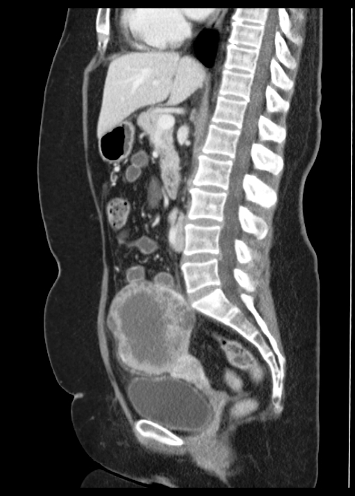
The patient is a 55-year-old para 1 female who presented with abdominal pain and a palpable mass. She had a history of inflammatory breast cancer two years prior to presentation. She underwent initial chemotherapy followed by radical mastectomy and lymphadenectomy revealing metastatic disease in her lymph nodes. She also received chest wall radiotherapy. Later she developed pain in her lower abdomen and palpated a mass. A CT scan revealed a 10 cm complex mass arising from the right ovary (Fig 1). CA-125 was markedly elevated at 1370 U/ml.
Her family history was remarkable for a sister who had breast cancer, female cousin who had ovarian cancer, and her mother died of uterine cancer.
Subsequently she underwent total abdominal hysterectomy and bilateral salpingo-oopherectomy with omentectomy and right pelvic lymphadenectomy. Operation findings included a right adnexal tumor with dense adhesions.
Gross:
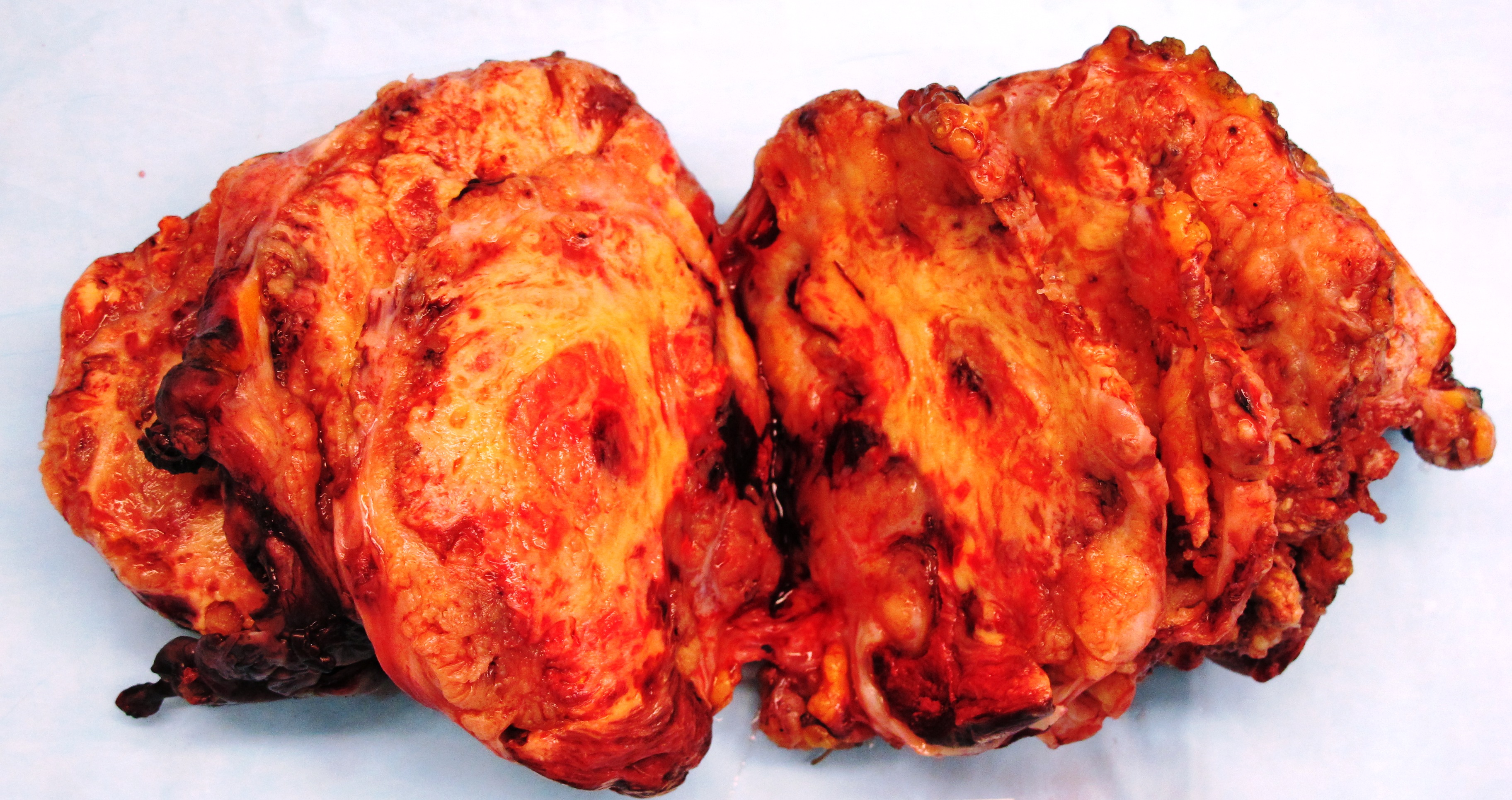
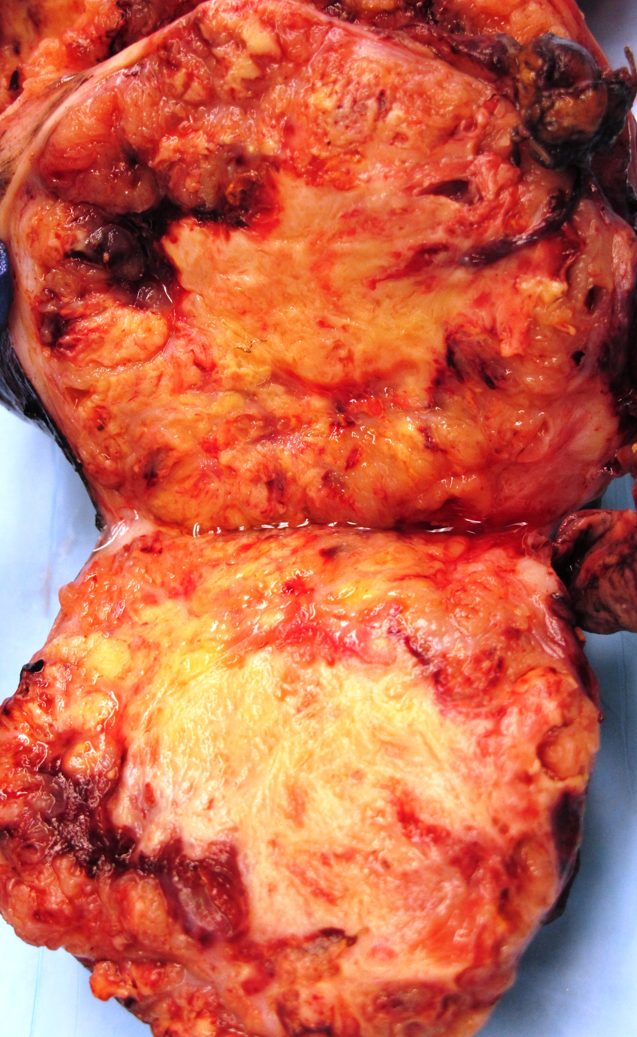
Received was a 419 gram previously disrupted ovarian mass (11.0 x 10.5 x 8.0 cm). The mass is oval, slightly nodular and is predominantly (approximately 80%) covered with a gray-pink smooth membrane with blotchy hemorrhage and without excrescences. Cut surfaces reveal heterogeneous, lobulated yellow to brown areas intermixed with hemorrhage and necrosis. Rare viable tumor areas are light-brown, soft and nodular (Fig 2, 3).
Images:
 |
 |
 |
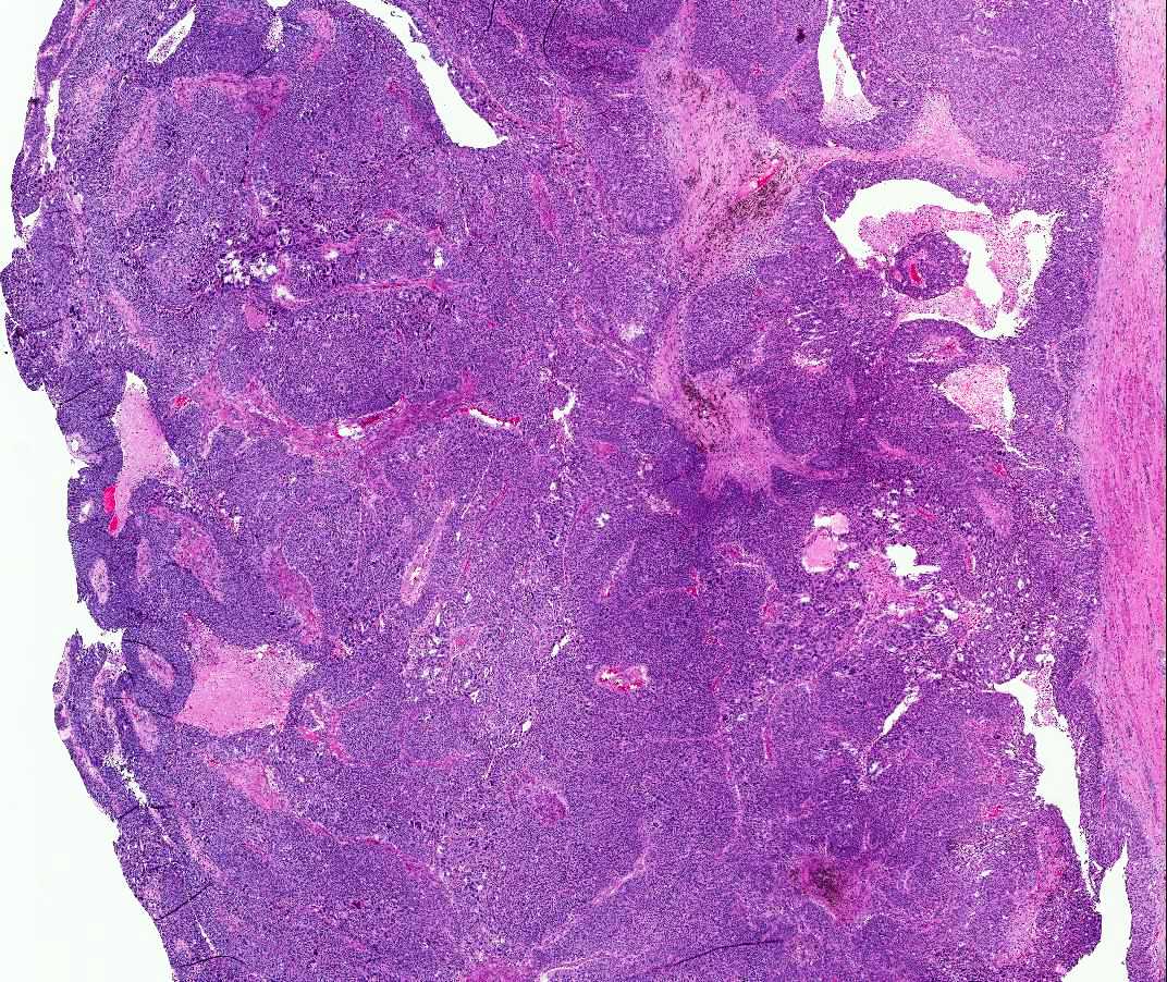
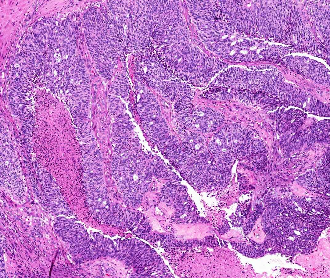
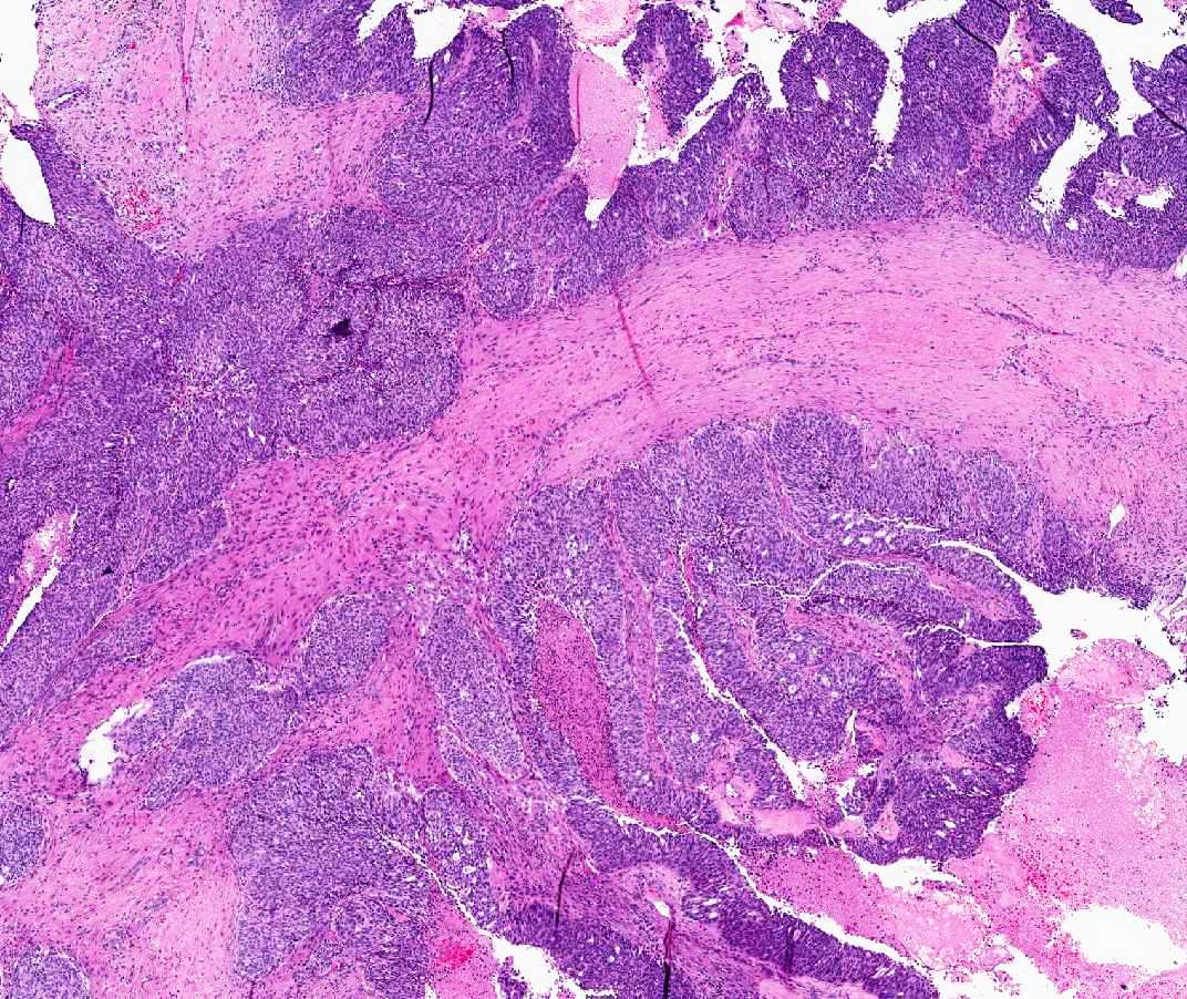
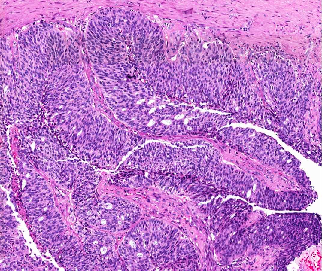
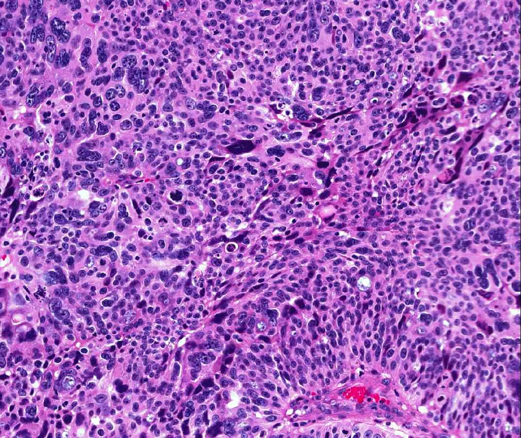
Microscopic photographs:
 |
 |
 |
||
 |
 |
 Meet our Residency Program Director
 LeShelle May
 Chancellor Gary May