Residency Program - Case of the Month
August 2013 - Presented by Sarah Barnhard, M.D.
Clinical History:
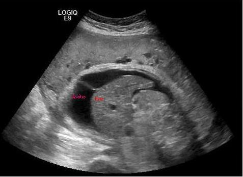
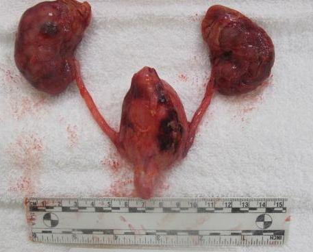
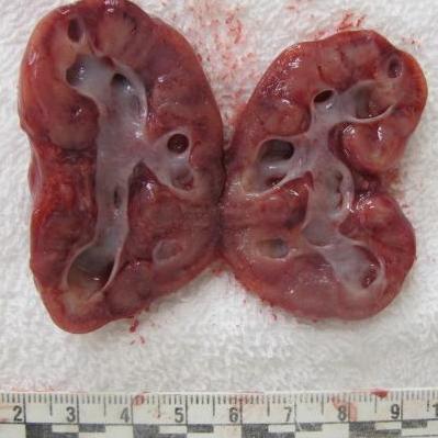
A 22 year old G1P0 female with an uncomplicated pregnancy history was at 31 weeks 6/7 days gestational age by last menstrual cycle when she presented to her OB clinic for routine screening. Her uterine size was significantly less than expected for gestational age, and thus an ultrasound at an outside hospital was performed. There was imaging concern for congenital anomalies, so she was transferred to our institution for management. Repeat fetal ultrasound showed severe oligohydramnios, marked ascites (image 1), bilateral hydronephrosis, a markedly distended thick-walled bladder, and a narrow thoracic diameter suggestive of hypoplastic lungs. Fetal echocardiography revealed a constricted foramen ovale and right sided atrial and ventricular dilatation with concern for pulmonary hypertension.
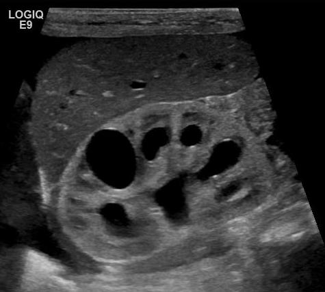
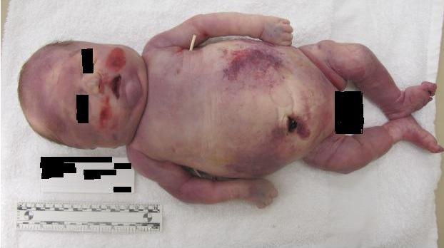
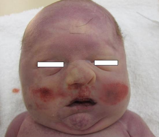
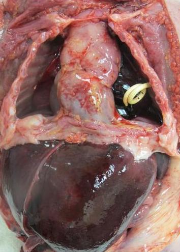
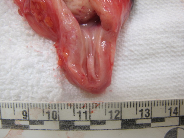
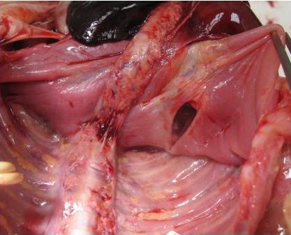
A male neonate was delivered by Caesarian section following fetal paracentesis intraoperatively. Post-delivery neonatal renal ultrasound revealed bilateral hydronephrosis (Image 2), and a distended bladder and proximal urethra with a “key-hole” morphology (Image 3). The neonate developed bilateral pneumothorax, severe refractory hypotension, and DIC with pulmonary hemorrhage. After multiple episodes of desaturation, the decedent expired approximately 21 hours after delivery. An autopsy was performed (images below).
Images:


 Meet our Residency Program Director
 LeShelle May
 Chancellor Gary May