
Residency Program - Case of the Month
April 2016 - Dr. Andrew Jones & Dr. Karen Matsukuma
History
The patient is a 65-year-old woman with a history of hypothyroidism, colonic tubular adenomas (including a 1.3 cm tubulovillous adenoma with high grade dysplasia, removed several years before), and new onset hematochezia. She underwent a colonoscopy two months prior to presentation as part of her hematochezia workup, which revealed a 1 cm firm and rubbery rectal mass, in addition to other adenomatous polyps. The mass was less than 1cm from the anal verge. The mass was biopsied, and the biopsy fragments were submitted for histologic examination.
On transrectal ultrasound, no evidence of submucosal infiltration of the lesion was identified. However, a subsequent PET scan demonstrated increased activity in one mesorectal lymph node; MRI demonstrated no metastatic lesions to the brain.
Gross Findings
Grossly, the tissue fragments measured 0.3 cm in greatest dimension and were tan-brown and otherwise unremarkable GI biopsies.
Microscopic Findings
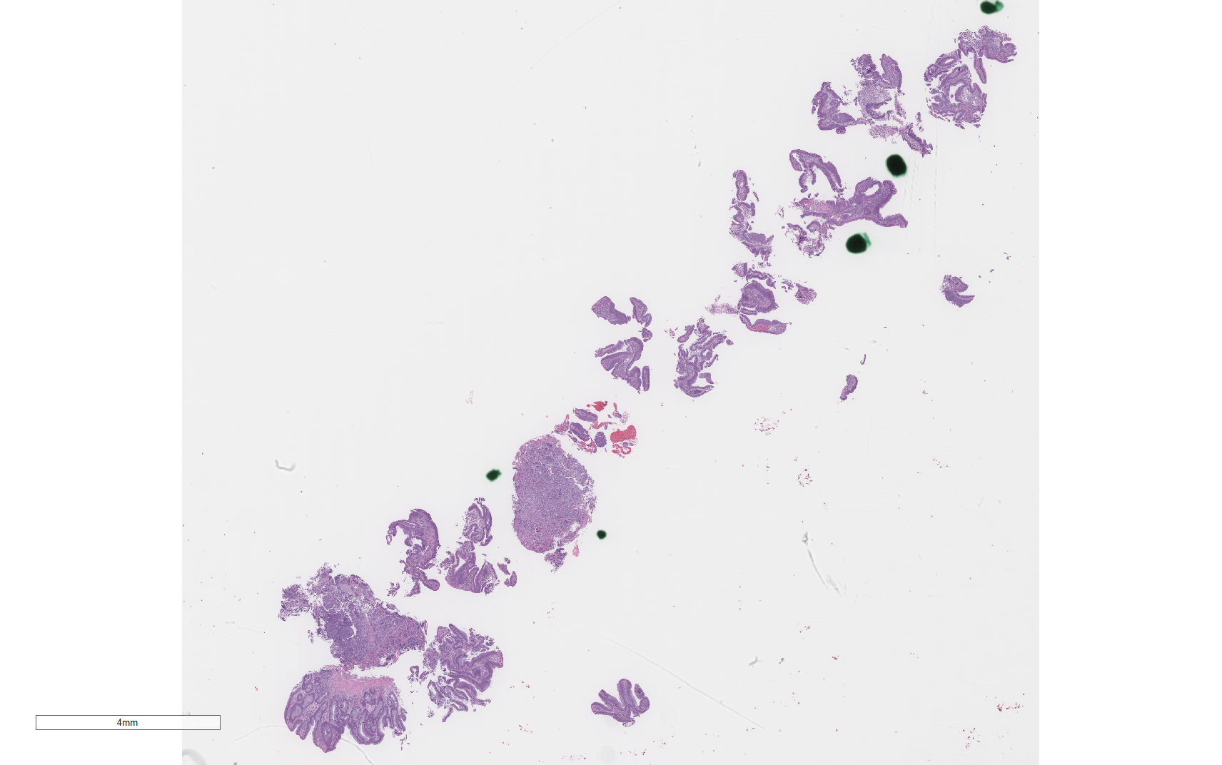
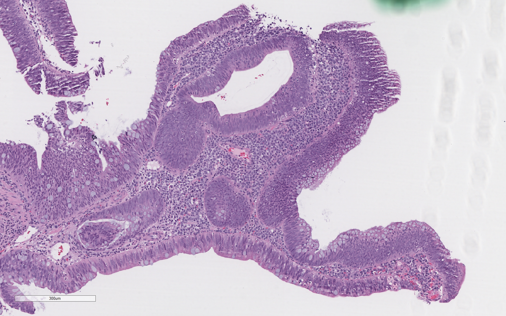
Histologic sections demonstrate multiple fragments of colonic mucosa (Figure 1). The epithelium was composed of densely packed pseudostratified columnar cells with hyperchromatic, pencillate nuclei and numerous mitotic figures (Figure 2). No necrosis, cribriforming, or significant nuclear pleomorphism was observed; areas of villous architecture were noted.
| Figure 1 | Figure 2 | |
 |
 |
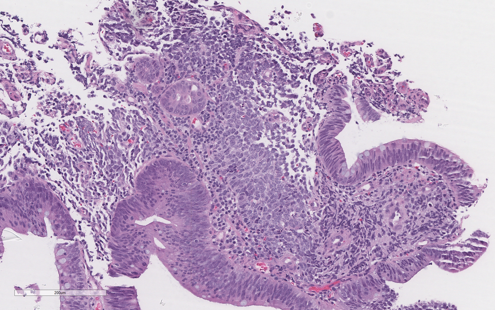
Additionally, the included fragments of lamina propria showed sheets and nests of medium-sized blue cells with minimal cytoplasm and large nuclei with variably prominent nucleoli (Figures 3 & 4).
| Figure 3 | Figure 4 | |
 |
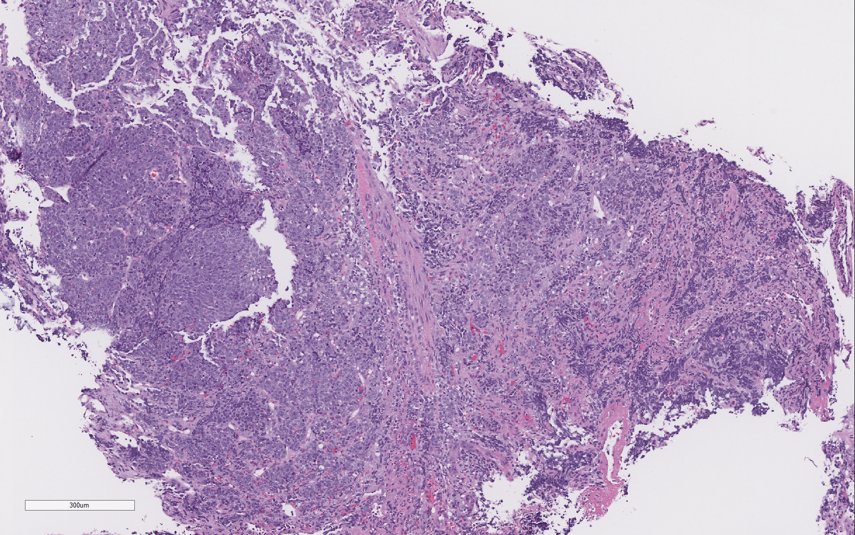 |
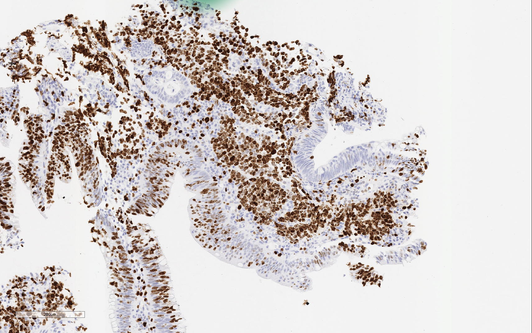
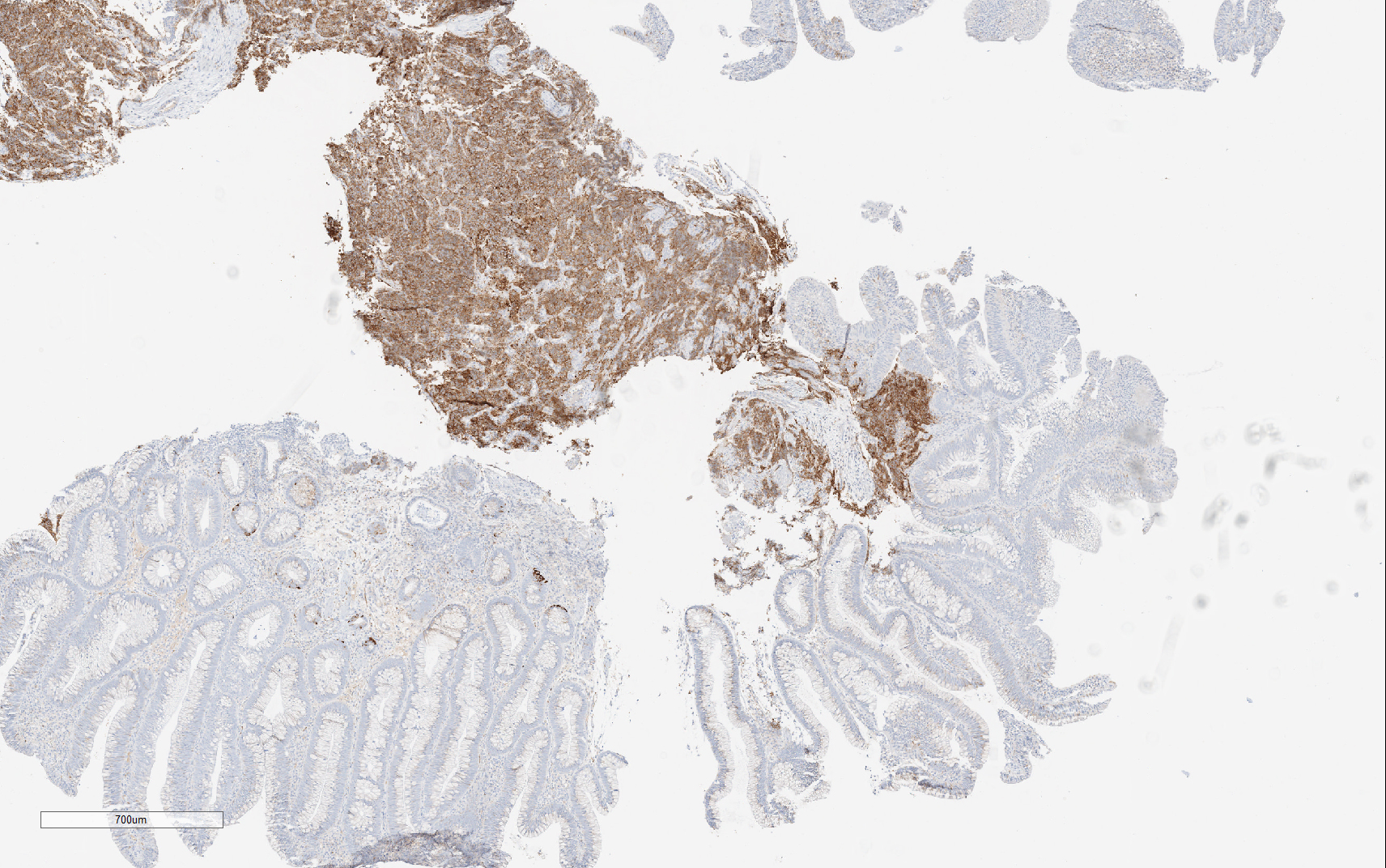
Numerous mitotic figures were observed; no necrosis was present. Immunohistochemical staining with Ki-67 revealed a proliferative index of 90% (Figure 5). The cells in the lamina propria were also positive for synaptophysin (Figure 6) and CAM5.2 and negative for chromogranin, CDX2, LCA, WT1, p63, CK7, CK20, and MART-1.
| Figure 5 | Figure 6 | |
 |
 |
What is the diagnosis?
Choose one answer and submit.
C. Combined neuroendocrine carcinoma (G3) and tubulovillous adenoma.
> Learn more about this diagnosis.
 Meet our Residency Program Director
 LeShelle May
 Chancellor Gary May