
Residency Program - Case of the Month
June 2016 - Presented by Dr. Saba Ali & Dr. Mingyi Chen
Clinical History:
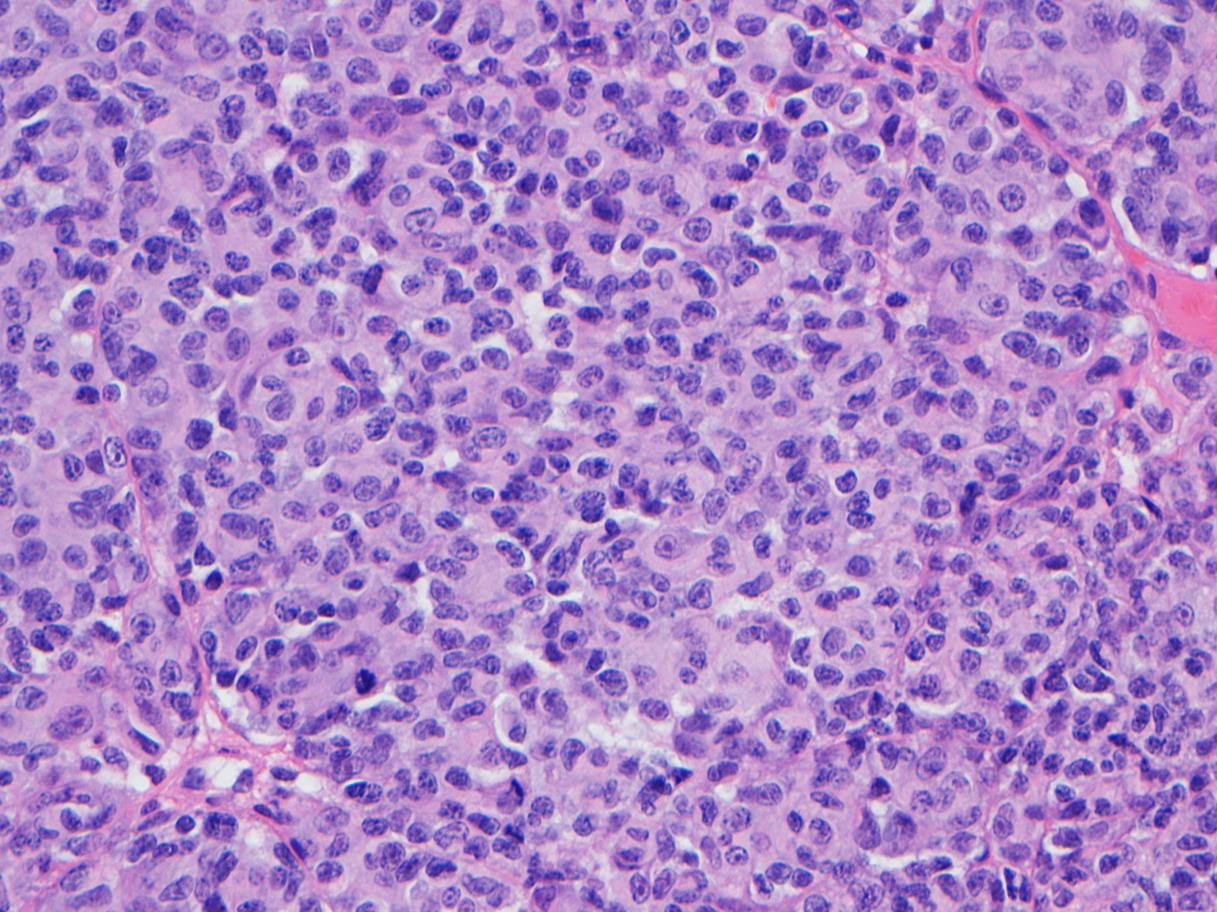
A 62-year-old female with a remote past medical history of hairy cell leukemia (HCL) presented to an outside hospital with progressively worsening headaches accompanied by short term memory loss, expressive aphasia, and lethargy. Subsequent brain imaging to rule out stroke showed significant brain metastases characterized by three parenchymal masses with vasogenic edema. She was transported to UC Davis Medical Center for further care. Additional imaging studies showed multiple nodules in the right middle lobe of the lung and spleen with subcarinal lymphadenopathy. An excisional biopsy of the left frontal brain mass was performed.
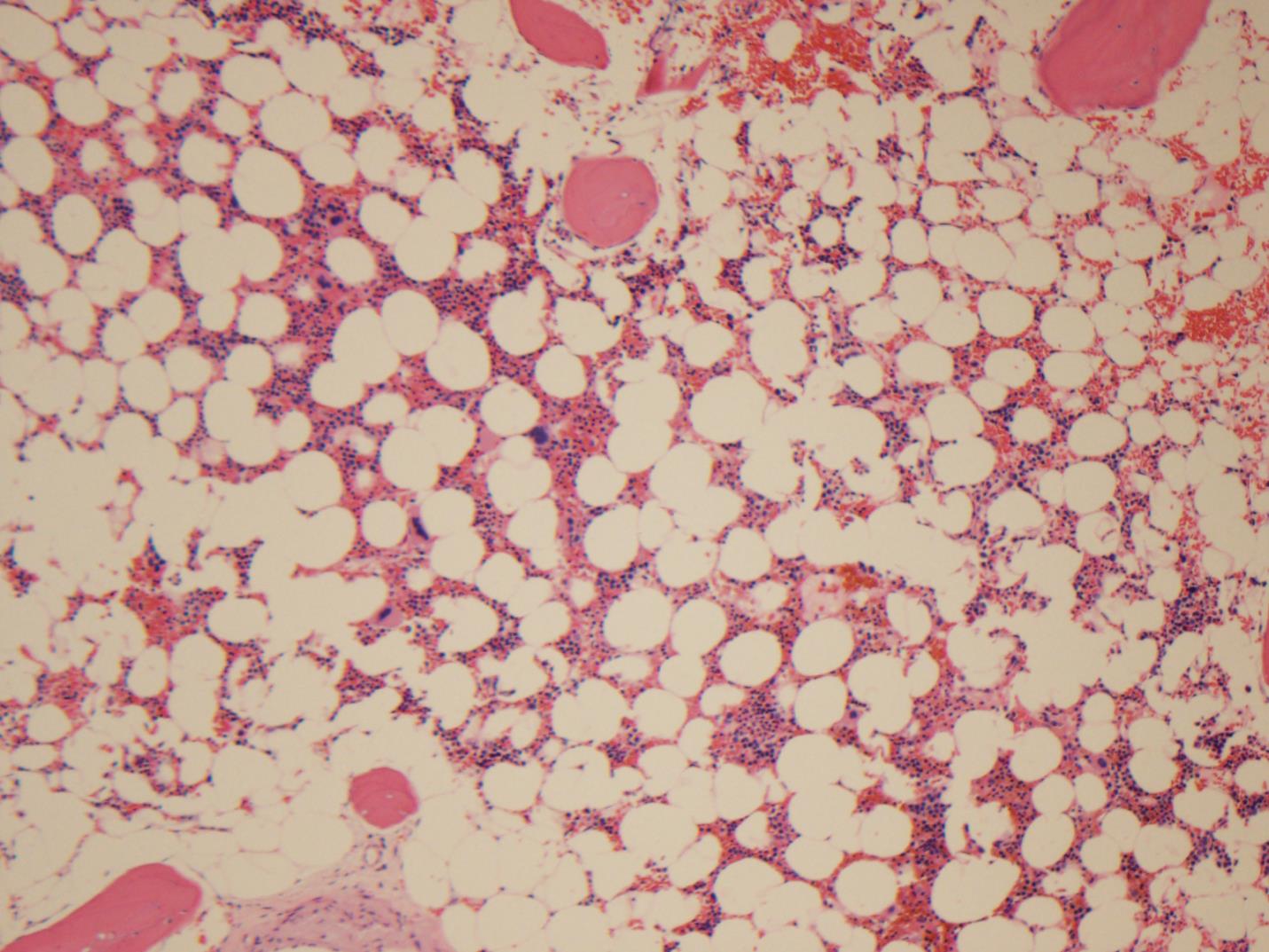
Additionally at this time, blood work up showed persistent pancytopenia with monocytopenia. Her initial diagnosis of HCL was made in 1993 when she presented with cytopenias, malaise, and splenomegaly. Given this history, a bone marrow biopsy was performed.
Microscopic Images:
 |
 |
|
| Figure 1 - Excisional biopsy of left frontal brain mass (H&E, 40x) |
Figure 2 - Peripheral blood |
|
 |
 |
|
| Figure 3 - Bone marrow biopsy (H&E, 40x) |
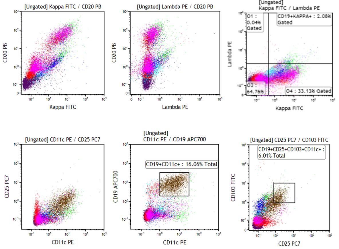
Figure 4 - Flow cytometry of |
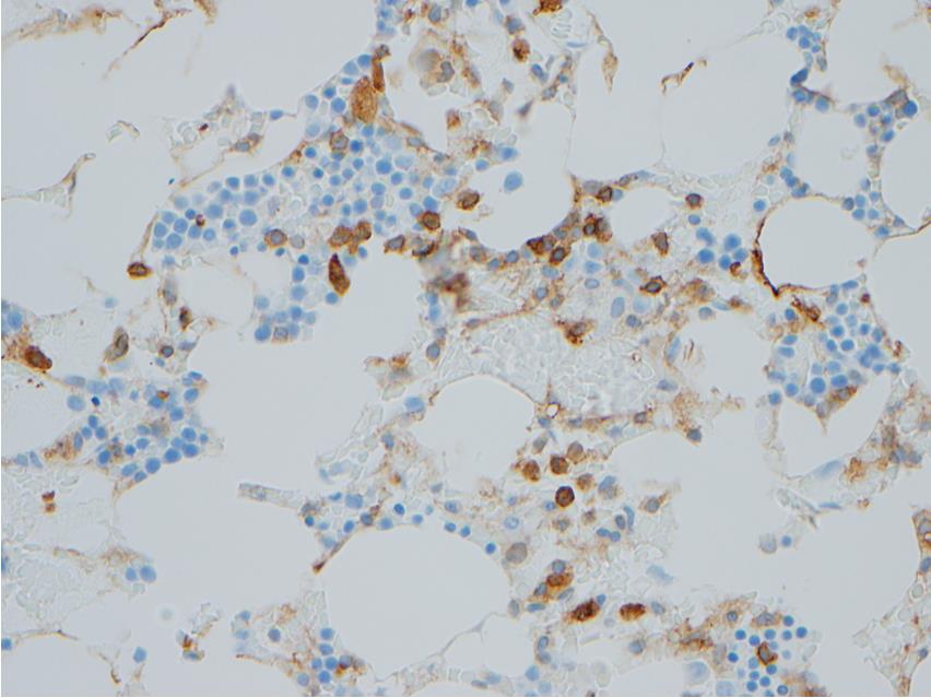
Immunohistochemistry:
 |
 |
|
| Figure 5 - Left brain frontal mass, HMB-45 (IHC). |
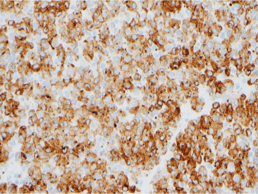
Figure 6 - Bone marrow biopsy, |
What gene is most commonly found in hairy cell leukemia and also associated with metastatic melanoma?
Choose one answer and submit.
 Meet our Residency Program Director
 LeShelle May
 Chancellor Gary May