
Residency Program - Case of the Month
May 2017 - Presented by Dr. Trevor Starnes & Dr. Tao Wang
Clinical History
The patient was a 21-month-old, previously healthy girl who presented with abdominal distension, emesis, and irritability to an outside hospital. She was found to have a liver mass. A liver biopsy was performed under anesthesia during which she developed bronchospasm. She was then intubated on propofol for 48 hours, developed propofol infusion syndrome, and later succumbed to complications of propofol infusion syndrome. An autopsy was performed.
Gross Photographs
Click image to enlarge
|
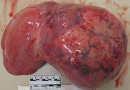
Figure 1 |
 |
|
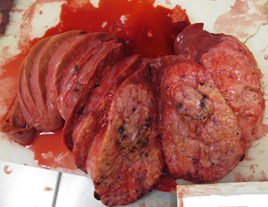
Figure 2 |
 |
Additional Relevant Gross Findings
The lungs also contained several subpleural white nodules ranging from 2-3 mm on the left upper and right middle lobes.
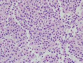
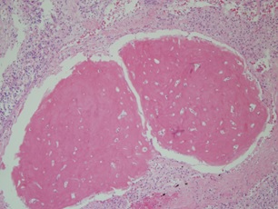
Microscopic Description
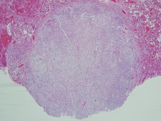
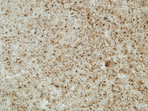
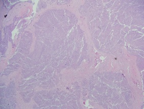
The liver mass lacks normal liver lobular architecture and does not contain bile ducts. Thick bands of collagen run through the mass. Between the bands of collagen are small, immature hepatocytes. The hepatocytes have minimal pleomorphism and do not have a substantially increased N:C ratio. There are large areas of hemorrhage and necrosis. There are focal areas of osteoblastic cells with osteoid. The uninvolved liver is unremarkable.
Click on image to enlarge
|
Figure 3 |
|
|
Figure 4 |
 |
|
Figure 5 |
 |
|
Figure 6 |
 |
|
Figure 7 |
 |
|
Figure 8 |
 |
|
Figure 9 |
 |
What is the diagnosis of the Liver Mass?
Choose one answer and submit.
B. Hepatoblastoma, mixed epithelial and mesenchymal type
There is also metastatic disease to the lungs.
> Learn more about this diagnosis.
 Meet our Residency Program Director
 LeShelle May
 Chancellor Gary May