
Residency Program - Case of the Month
March 2018 - Presented by Dr. Trevor Starnes (Mentored by: Dr. Karen Matsukuma)
Clinical History
The patient is a 64-year-old woman who presented to an outside gastroenterologist for evaluation of dysphagia. During esophagogastroduodenoscopy, the patient was noted to have an submucosal mass in the duodenum, possibly representing a prominent papilla. Subsequent magnetic resonance cholangiopancreatography did not reveal an ampullary mass, and liver function tests were unremarkable.
Microscopic Description
Sections of the 1.3 cm mass demonstrate a submucosal neoplasm beneath unremarkable duodenal mucosa. The neoplasm is composed of a combination of epithelioid cells in nests (Figures 1-3) with spindled cells (Figures 1 and 2) and scattered ganglion cells (Figure 3). The epithelioid cells are round with central nuclei and salt-and-pepper chromatin. The spindled cells are bland, form broad fascicles, and encircle the nests of epithelioid cells. Ganglion cells are large with prominent nucleoli and eosinophilic cytoplasm. S100 highlights the spindled cells (Figure 4); HMB-45 is negative (Figure 6). Synaptophysin is positive in the epithelioid cells (Figure 5), and the Ki-67 of these cells is low (Figure 7).
Click on image to enlarge.
Figure 1: Low power view of the duodenal neoplasm, 40x
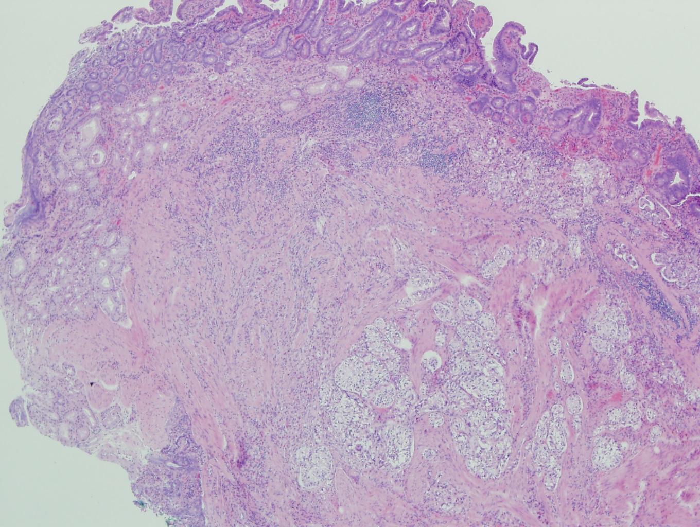
Figure 2: The duodenal neoplasm has spindled and epithelioid cells, 200x
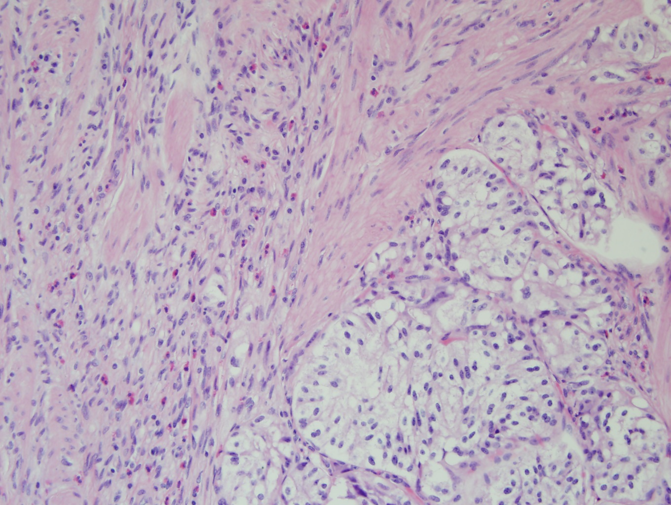
Figure 3: Among the epithelioid cells are scattered large cells with prominent nucleoli, 400x
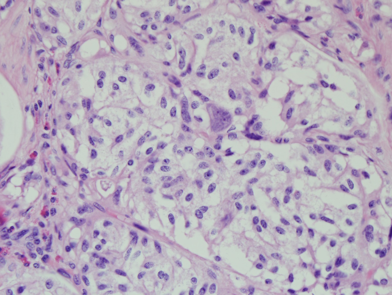
Figure 4: Staining of spindled cells in the duofenal neoplasm, 100x
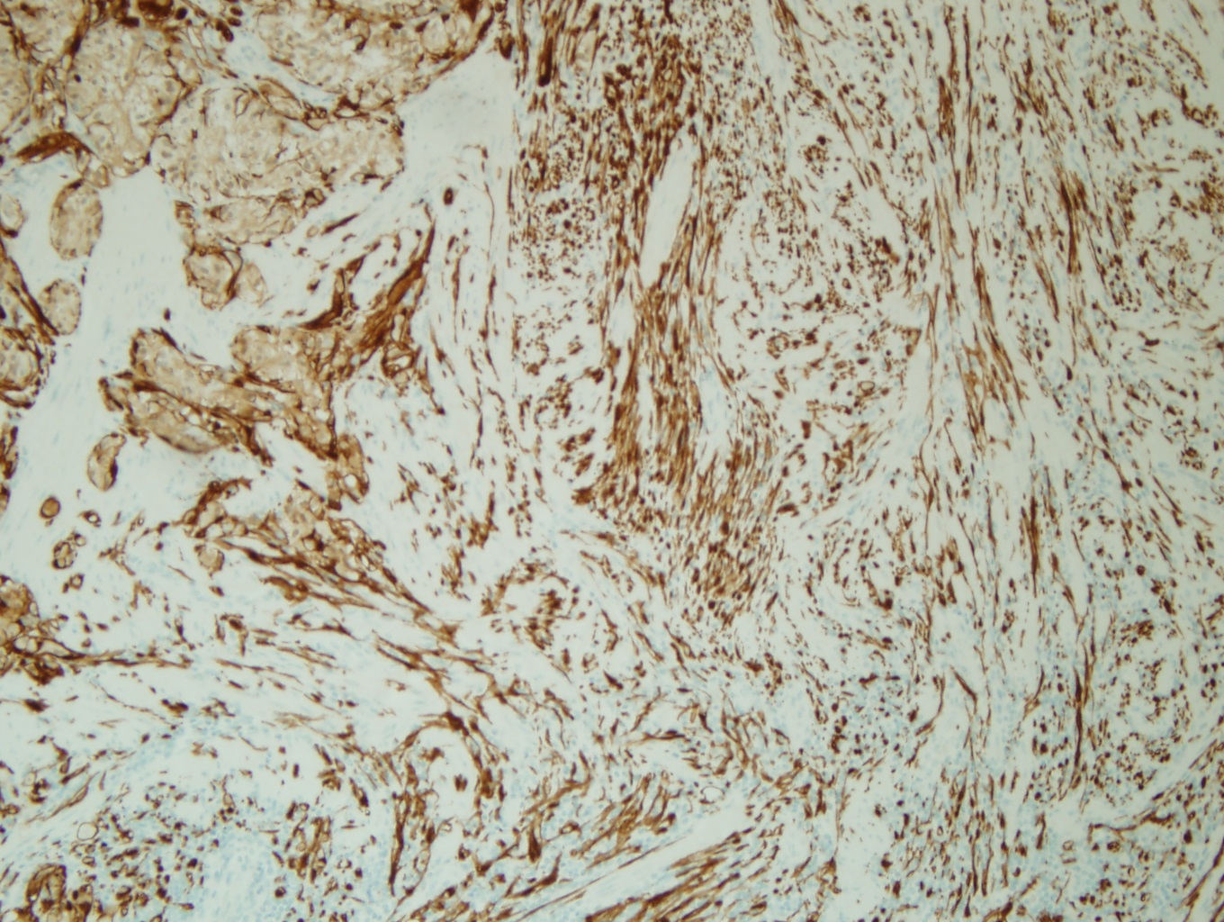
Figure 5: Synaptophysin staining of epithelioid and large cells in the duodenal neoplasm, 100x
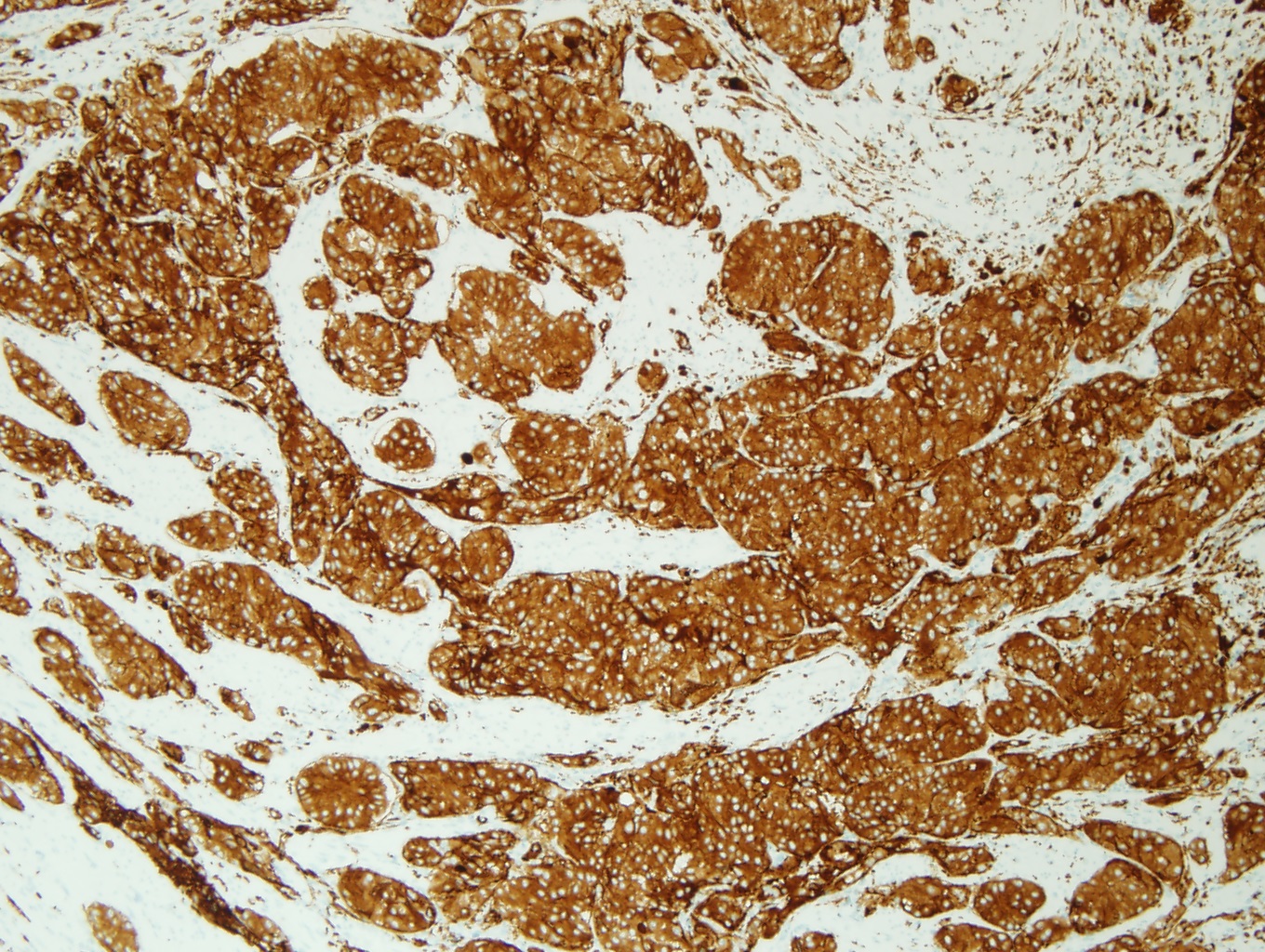
Figure 6: HMB-45 is negative in the duodenal neoplasm, 100x
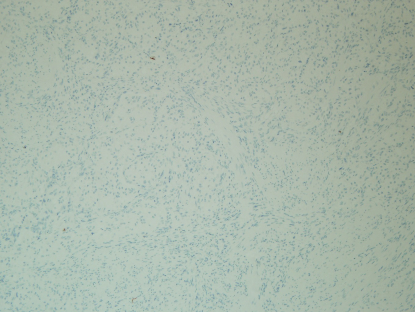
Figure 7: Ki-67 shows a low mitotic rate in the neoplastic cells, 100x

What is the diagnosis?
Choose one answer and submit.
 Meet our Residency Program Director
 LeShelle May
 Chancellor Gary May