
Resident Program - Case of the Month
February 2019 - Presented by Dr. Ryan Thomas (Mentored by Dr. Anthony Karnezis)
Clinical History
A 29-year-old G5P2022 presented for repeat cesarean section at 38 weeks gestation. The pregnancy was a twin gestation complicated by intrauterine fetal demise of one twin at 11 weeks gestation. Subsequent chorionic villous sampling of both twins showed a normal 46 XX karyotype in the living twin and a 69 XXX genotype in the demised twin. The pregnancy was maintained with close monitoring and the patient delivered a viable 2740 g female infant via cesarean section. The placenta was sent for pathologic examination.
Pathological Findings
Gross examination of the placenta of the viable twin was unremarkable. The placenta of the demised twin revealed a thickened and fibrotic fetal placental surface with an underlying area of hydropic villi as well as a 3.4 x 2.6 x 0.2 cm flat, ovoid, firm rubbery plaque with punctate brown pigmentation within the placental membrane (Figure 1).
Click on images to enlarge.
| Figure 1 |
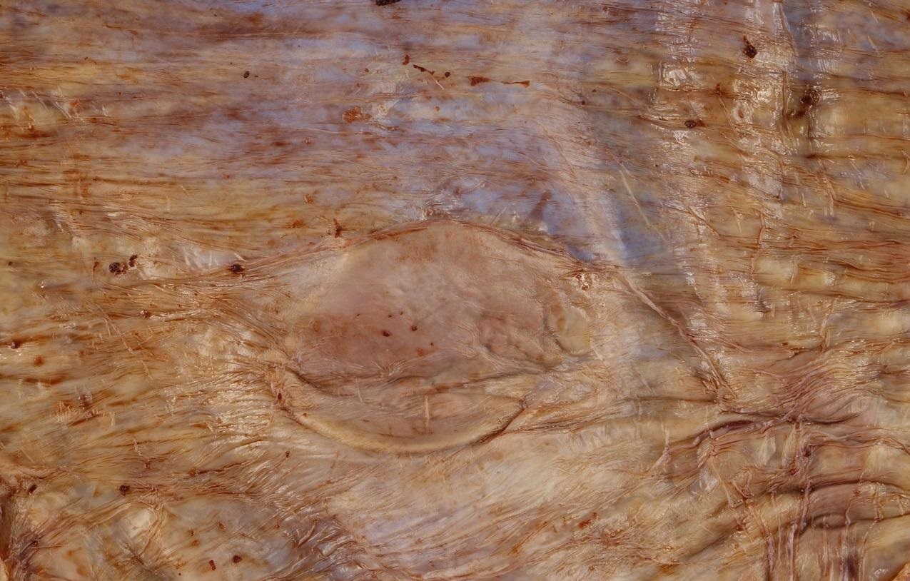 |
The placental membranes of the demised twin were remarkable for a 3.4 x 2.6 x 0.2 cm ovoid, firm rubbery plaque.
Microscopic examination of the abnormal placental disc revealed large hydropic villi admixed with small fibrotic villi (Figure 2). Microscopic examination of the placental membrane plaque revealed symmetrical immature cartilage formation amidst necrotic debris with mineralization (Figure 3).
| Figure 2 |
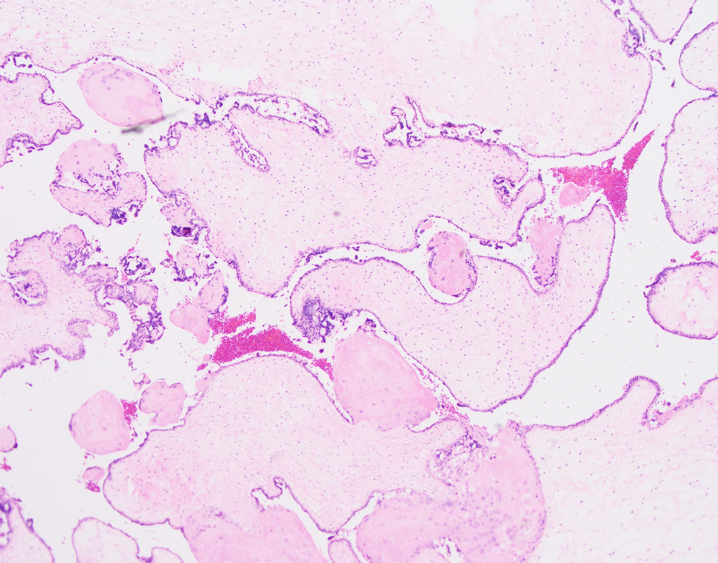 |
This image shows the large hydropic villi seen on a section from the fibrotic placental disc (40X magnification).
| Figure 3 |
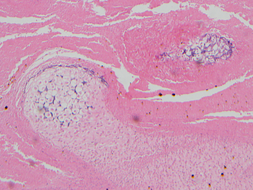 |
Notice the immature cartilage formation as well as mineralization seen on this section of the membrane plaque. (100X magnification).
What is the most likely diagnosis for the membrane abnormality?
Choose one answer and submit.
 Meet our Residency Program Director
 LeShelle May
 Chancellor Gary May